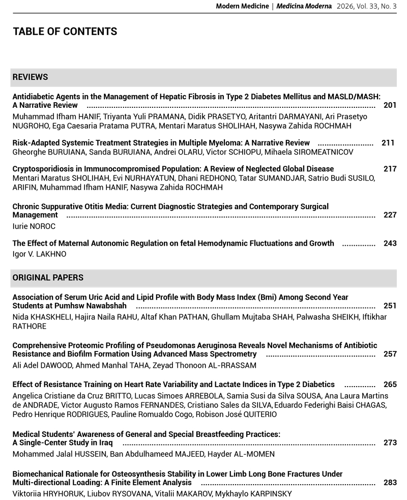
Background. In the general structure of trauma, fractures of the bones of the lower extremities account for 47.3%, among which diaphyseal injuries occupy the leading place. The choice of the optimal type of fixator (osseous, intramedullary, or external) remains a subject of discussion, since the stability of the “bone – implant” system directly determines the nature of the regenerative process and the timing of consolidation.
Objective: based on three-dimensional mathematical modeling using the finite element method, to conduct a comparative analysis of the stress-strain state of femur and tibia models with fractures at different diaphyseal levels under four variants of multidirectional loads when using four common osteosynthesis systems.
Materials and methods. Three-dimensional finite element models of the femur and tibia with transverse diaphyseal fractures in the upper, middle, and lower thirds were constructed. The stability of fixation with a bone plate, Ilizarov external fixator, unilateral rod fixator, and interlocking intramedullary nailing (IMN) was investigated. Four loading options were modeled: Option 1 (pure axial compression), Option 2 (axial compression and frontal bending), Option 3 (axial compression and sagittal bending), Option 4 (axial compression and torsion). Results. Bone osteosynthesis provides absolute rigidity (Δ = 0.00 mm) in the 3rd and 4th segments of both bones, but in low fractures of the tibia (n/3), stresses increase to 6.70 MPa, and micromobility – to 0.30 mm. The spike apparatus demonstrates universal isotropic elasticity (Δ = 0.01–0.05 mm) in both segments under all types of loads. The rod apparatus in n/3 fractures reveals critical instability: stresses on the femoral rods reach 38.2 MPa, and the displacement of the tibia fragments is 1.90 mm. The IMN method is the standard for the hip at all levels (Δ ≤ 0.15 mm); however, in low fractures of the tibia (n/3) under combined forces, fragment displacement reaches extraphysiological limits of 1.42–1.60 mm.
Conclusions. The stability of the bone-implant system is determined by the design features of the fixation device and the extent of diaphyseal damage. None of the osteosynthesis methods studied offers absolute mechanical versatility across all bone segments. Intramedullary osteosynthesis is the optimal method, except for fractures in the distal third of the tibial shaft, where a wire-based external fixation device proved more biomechanically stable.